")
Mastering Jugular Venous Pressure (JVP) Measurement in Clinical Cardiology: Why This Classic Bedside Assessment Remains Indispensable in Modern Practice. Discover the Latest Techniques, Clinical Impact, and Future Innovations. (2025)
- Introduction: The Enduring Relevance of JVP in Cardiology
- Anatomy and Physiology of the Jugular Venous System
- Clinical Techniques for Accurate JVP Measurement
- Common Pitfalls and How to Avoid Them
- JVP in the Diagnosis and Management of Heart Failure
- Technological Advances: Digital and Ultrasound-Assisted JVP Assessment
- Comparative Effectiveness: JVP Versus Other Hemodynamic Markers
- Training, Competency, and Standardization in JVP Measurement
- Public and Professional Awareness: Trends and Forecasts (Estimated 15% Growth in Clinical Adoption by 2030)
- Future Directions: AI, Remote Monitoring, and the Evolving Role of JVP in Cardiology
- Sources & References
Introduction: The Enduring Relevance of JVP in Cardiology
The measurement of jugular venous pressure (JVP) remains a cornerstone of bedside cardiovascular assessment, retaining its clinical relevance well into 2025. JVP reflects the pressure within the right atrium and central venous system, providing a non-invasive window into cardiac function, particularly right-sided heart hemodynamics. Despite the proliferation of advanced imaging modalities and laboratory diagnostics, the physical examination of JVP continues to offer immediate, cost-effective, and valuable information for clinicians evaluating patients with suspected or established cardiovascular disease.
JVP assessment is especially critical in the diagnosis and management of conditions such as heart failure, constrictive pericarditis, cardiac tamponade, and volume overload states. By observing the height and waveform of the jugular venous pulse, clinicians can infer right atrial pressure, estimate central venous pressure, and detect abnormal cardiac filling patterns. This bedside skill is emphasized in clinical guidelines and remains a fundamental component of the cardiovascular examination taught in medical education worldwide.
The enduring importance of JVP measurement is underscored by its inclusion in the recommendations of leading cardiology organizations. For example, the American College of Cardiology and the European Society of Cardiology both highlight the role of JVP assessment in the evaluation of heart failure and other cardiac conditions. These organizations, recognized as global authorities in cardiovascular medicine, advocate for the integration of JVP measurement with other clinical findings to guide diagnosis, risk stratification, and therapeutic decision-making.
In an era marked by rapid technological advancement, the value of JVP measurement lies in its accessibility and immediacy. Unlike echocardiography or invasive hemodynamic monitoring, JVP assessment requires no specialized equipment and can be performed at the bedside, in outpatient clinics, or even in resource-limited settings. This makes it an indispensable tool for clinicians worldwide, ensuring that critical information about a patient’s volume status and cardiac function can be obtained promptly and efficiently.
As the field of cardiology continues to evolve, the measurement of JVP stands as a testament to the enduring utility of clinical examination skills. Its continued relevance in 2025 reflects both its diagnostic power and its adaptability alongside modern diagnostic technologies, reinforcing the importance of a thorough and nuanced bedside assessment in the care of patients with cardiovascular disease.
Anatomy and Physiology of the Jugular Venous System
The jugular venous system plays a pivotal role in the assessment of central venous pressure, which is a key parameter in clinical cardiology. The jugular veins, primarily the internal jugular vein (IJV) and the external jugular vein (EJV), are responsible for draining deoxygenated blood from the brain, face, and neck into the superior vena cava, and ultimately into the right atrium of the heart. The IJV is the preferred site for clinical evaluation of jugular venous pressure (JVP) due to its direct anatomical continuity with the right atrium and its relative protection from external compression.
Anatomically, the IJV runs deep to the sternocleidomastoid muscle, beginning at the jugular foramen at the base of the skull and descending to join the subclavian vein behind the clavicle. The EJV, in contrast, is more superficial and less reliable for JVP assessment because it is more susceptible to external pressure and anatomical variation. The right IJV is typically used for JVP measurement, as it provides a more direct, unobstructed pathway to the right atrium, minimizing the influence of thoracic structures.
Physiologically, the jugular venous pulse reflects the dynamic changes in right atrial pressure throughout the cardiac cycle. The waveform observed in the IJV consists of several distinct components: the ‘a’ wave (atrial contraction), ‘c’ wave (bulging of the tricuspid valve during ventricular contraction), ‘x’ descent (atrial relaxation), ‘v’ wave (venous filling of the right atrium), and ‘y’ descent (opening of the tricuspid valve and rapid ventricular filling). These waveforms provide valuable insights into right heart function and can indicate the presence of specific cardiac pathologies, such as tricuspid regurgitation, right heart failure, or pericardial disease.
The measurement of JVP is a non-invasive bedside technique that estimates central venous pressure by assessing the vertical height of venous pulsation above the sternal angle. Accurate interpretation requires a thorough understanding of the jugular venous anatomy and the physiological basis of the venous pulse. JVP assessment remains a cornerstone of cardiovascular examination, aiding in the diagnosis and management of conditions such as heart failure, volume overload, and constrictive pericarditis.
The importance of JVP measurement is underscored by its inclusion in clinical guidelines and educational resources from leading cardiology organizations, such as the American College of Cardiology and the European Society of Cardiology. These bodies emphasize the value of JVP as a simple yet powerful tool in the bedside evaluation of cardiac patients.
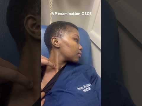
Clinical Techniques for Accurate JVP Measurement
Accurate measurement of Jugular Venous Pressure (JVP) is a cornerstone of bedside cardiovascular assessment, providing critical insights into right atrial pressure and overall hemodynamic status. The clinical technique for JVP measurement relies on visual inspection and anatomical landmarks, making it a skill that requires both knowledge and practice for reliable results.
The patient should be positioned at a 30 to 45-degree angle, with the head turned slightly away from the side being examined. This position optimizes visualization of the internal jugular vein, which is preferred over the external jugular vein due to its direct anatomical connection to the right atrium and reduced susceptibility to external pressure. Adequate lighting is essential, and the examiner should look for the characteristic double waveform pulsation of the internal jugular vein, distinguishing it from the carotid artery, which has a single, palpable pulse.
To measure JVP, the vertical distance between the highest point of venous pulsation and the sternal angle (Angle of Louis) is determined. The sternal angle is a reliable anatomical reference point, approximately 5 cm above the mid-right atrium in most adults. The measured vertical height (in centimeters) is added to this 5 cm reference to estimate the central venous pressure. A JVP greater than 3-4 cm above the sternal angle (or 8-9 cm above the right atrium) is generally considered elevated and may indicate conditions such as right heart failure, fluid overload, or constrictive pericarditis.
Several maneuvers can enhance the accuracy of JVP assessment. The hepatojugular reflux test, for example, involves applying gentle pressure over the liver to transiently increase venous return, which can accentuate jugular distension in cases of right-sided heart dysfunction. It is also important to differentiate true venous pulsations from carotid pulsations by observing for non-palpability, biphasic waveform, and changes with respiration or position.
While bedside JVP measurement is a non-invasive and cost-effective tool, it is subject to inter-observer variability and can be challenging in patients with obesity, short necks, or poor lighting. Training and repeated practice are essential for proficiency. In ambiguous cases, adjunctive methods such as ultrasound can be employed to visualize the internal jugular vein and improve diagnostic accuracy, as recommended by leading cardiology organizations such as the American College of Cardiology and the European Society of Cardiology. These bodies emphasize the continued relevance of JVP assessment as part of a comprehensive cardiovascular examination.
Common Pitfalls and How to Avoid Them
Accurate measurement of jugular venous pressure (JVP) is a cornerstone of bedside cardiovascular assessment, yet it is fraught with potential pitfalls that can compromise clinical decision-making. Recognizing and addressing these common errors is essential for clinicians to ensure reliable evaluation of central venous pressure and right heart function.
1. Misidentification of the Jugular Vein
A frequent error is mistaking the external jugular vein (EJV) for the internal jugular vein (IJV). The IJV is preferred for JVP assessment because it is more directly connected to the right atrium and is less likely to be affected by external factors such as muscle contraction or fascial planes. The EJV, being more superficial, can be misleading due to its variable anatomy and susceptibility to external pressure. To avoid this, clinicians should identify the IJV by its location—lateral to the carotid artery and deep to the sternocleidomastoid muscle—and by observing its characteristic undulating pulsation, which is non-palpable and varies with respiration and position.
2. Incorrect Patient Positioning
JVP should be measured with the patient reclined at a 30–45 degree angle. Too flat a position may obscure venous pulsations, while too upright a position may cause them to disappear below the clavicle. Adjusting the angle to best visualize the top of the venous column is recommended. The head should be turned slightly away from the side being examined, but not so much as to tense the neck muscles, which can obscure the vein.
3. Confusing Arterial and Venous Pulsations
Carotid arterial pulsations can be mistaken for venous pulsations. Unlike the carotid pulse, the jugular venous pulse is biphasic, non-palpable, and changes with respiration and abdominal pressure. Palpation and careful observation of waveform characteristics help distinguish between the two.
4. Inaccurate Reference Point
JVP is measured as the vertical distance above the sternal angle, which is approximately 5 cm above the right atrium in most adults. Using an incorrect reference point, such as the clavicle or the mid-axillary line, can lead to significant errors in estimation. Consistent use of the sternal angle is recommended by major cardiology guidelines, including those from the American College of Cardiology and the European Society of Cardiology.
5. Overlooking Physiological and Pathological Influences
Factors such as elevated intra-thoracic pressure, tricuspid regurgitation, or superior vena cava obstruction can alter JVP readings. Awareness of these conditions and correlating JVP findings with the overall clinical context is crucial for accurate interpretation.
By systematically addressing these pitfalls—through proper technique, anatomical knowledge, and clinical correlation—clinicians can enhance the reliability of JVP measurement, supporting more accurate diagnosis and management of cardiovascular conditions.
JVP in the Diagnosis and Management of Heart Failure
The assessment of jugular venous pressure (JVP) remains a cornerstone in the clinical evaluation of patients with suspected or established heart failure. JVP reflects right atrial pressure and, by extension, central venous pressure, providing valuable insights into a patient’s volume status and cardiac function. Accurate measurement and interpretation of JVP are essential for both the diagnosis and ongoing management of heart failure, as recommended by leading cardiology organizations such as the American College of Cardiology and the European Society of Cardiology.
In heart failure, elevated JVP is a classic physical finding indicating increased right-sided cardiac pressures, often due to fluid overload or impaired right ventricular function. Clinicians typically assess JVP by positioning the patient at a 30–45 degree angle and observing the vertical height of the internal jugular vein above the sternal angle. A JVP greater than 3–4 cm above the sternal angle is generally considered abnormal and suggests elevated central venous pressure. This finding, when combined with other clinical signs such as peripheral edema and pulmonary rales, strengthens the diagnosis of heart failure.
JVP measurement is particularly valuable in differentiating between cardiac and non-cardiac causes of dyspnea. For example, a normal JVP in a patient with shortness of breath may point toward a pulmonary etiology rather than heart failure. Conversely, a raised JVP supports the diagnosis of heart failure and can help distinguish between right- and left-sided failure, as well as between acute and chronic presentations.
Beyond diagnosis, serial JVP assessments are instrumental in guiding the management of heart failure. Monitoring JVP trends allows clinicians to evaluate the effectiveness of diuretic therapy and other interventions aimed at reducing volume overload. A decreasing JVP in response to treatment is a favorable sign, while persistently elevated JVP may prompt adjustments in therapy or further investigation for refractory heart failure or concomitant conditions such as tricuspid regurgitation or constrictive pericarditis.
Despite advances in imaging and hemodynamic monitoring, bedside JVP assessment remains a practical, non-invasive, and cost-effective tool. Its utility is emphasized in clinical guidelines from the American College of Cardiology and the European Society of Cardiology, which advocate for its routine use in the evaluation and follow-up of heart failure patients. Mastery of JVP measurement thus continues to be a fundamental skill in clinical cardiology, directly impacting patient outcomes in 2025 and beyond.
Technological Advances: Digital and Ultrasound-Assisted JVP Assessment
Technological advances have significantly transformed the assessment of jugular venous pressure (JVP) in clinical cardiology, moving beyond traditional bedside examination to incorporate digital and ultrasound-assisted methods. These innovations aim to enhance the accuracy, reproducibility, and objectivity of JVP measurement, which is a critical parameter in evaluating right heart function and diagnosing conditions such as heart failure.
Digital tools, including smartphone applications and wearable devices, are increasingly being developed to assist clinicians in JVP assessment. These technologies utilize high-resolution cameras and image processing algorithms to detect subtle changes in neck vein distension and pulsation. By providing real-time analysis and quantification, digital platforms can reduce observer variability and improve the consistency of JVP measurements. Some systems also integrate with electronic health records, facilitating longitudinal monitoring and telemedicine applications. The adoption of such digital solutions is supported by ongoing research and pilot programs in academic medical centers and hospitals worldwide, reflecting a broader trend toward digital health integration in cardiovascular care.
Ultrasound-assisted JVP assessment represents another major advancement. Point-of-care ultrasound (POCUS) enables direct visualization of the internal jugular vein, allowing clinicians to measure venous dimensions, assess collapsibility, and estimate central venous pressure with greater precision than visual inspection alone. Ultrasound guidance is particularly valuable in patients with challenging anatomy, obesity, or equivocal physical findings. Studies have demonstrated that ultrasound-based JVP estimation correlates well with invasive hemodynamic measurements, supporting its use as a noninvasive alternative in both inpatient and outpatient settings. The technique is now incorporated into training curricula and clinical guidelines by leading organizations such as the American College of Cardiology and the European Society of Cardiology, both of which are globally recognized authorities in cardiovascular medicine.
The integration of digital and ultrasound technologies into routine practice is not without challenges. Barriers include the need for clinician training, standardization of measurement protocols, and ensuring equitable access to advanced devices. Nevertheless, these innovations are poised to enhance the diagnostic utility of JVP assessment, supporting earlier detection of cardiac dysfunction and more personalized patient management. As technology continues to evolve, further improvements in automation, artificial intelligence, and remote monitoring are anticipated, reinforcing the central role of JVP measurement in modern cardiology.
Comparative Effectiveness: JVP Versus Other Hemodynamic Markers
The assessment of jugular venous pressure (JVP) remains a cornerstone of bedside clinical evaluation in cardiology, particularly for estimating right atrial pressure and diagnosing conditions such as heart failure. However, its comparative effectiveness against other hemodynamic markers—such as central venous pressure (CVP), pulmonary capillary wedge pressure (PCWP), and natriuretic peptide levels—has been the subject of ongoing research and debate.
JVP measurement offers a non-invasive, rapid, and cost-effective means of assessing central venous pressure. When performed correctly, it provides valuable information about a patient’s volume status and right heart function. Studies have shown that elevated JVP correlates with increased right atrial pressure and can predict adverse outcomes in heart failure patients. However, the accuracy of JVP assessment is highly operator-dependent and can be limited by patient factors such as obesity, neck anatomy, and lighting conditions.
In contrast, direct measurement of CVP via central venous catheterization is considered the gold standard for right atrial pressure assessment. While CVP provides precise quantitative data, it is invasive, carries risks of infection and thrombosis, and is not always feasible outside intensive care settings. Similarly, PCWP, measured through pulmonary artery catheterization, offers detailed insights into left-sided heart pressures and is valuable in complex cases of heart failure or shock. However, its invasive nature and associated complications have led to a decline in routine use, with guidelines recommending it primarily for select critically ill patients.
Biomarkers such as B-type natriuretic peptide (BNP) and N-terminal proBNP (NT-proBNP) have emerged as important adjuncts in the evaluation of heart failure. These blood tests provide objective, reproducible data and are useful for diagnosis, risk stratification, and monitoring response to therapy. Nevertheless, they do not offer real-time hemodynamic information and can be influenced by factors such as renal function, age, and obesity.
Comparative studies suggest that while JVP assessment is less precise than invasive measurements, it remains clinically valuable, especially when combined with other physical findings and laboratory data. The American College of Cardiology and American Heart Association continue to endorse JVP evaluation as part of the standard physical examination in heart failure and volume assessment. Ultimately, the choice of hemodynamic marker should be individualized, balancing the need for accuracy, invasiveness, and clinical context.
Training, Competency, and Standardization in JVP Measurement
Accurate assessment of jugular venous pressure (JVP) remains a cornerstone of bedside cardiovascular examination, providing critical insights into right atrial pressure and overall hemodynamic status. However, the reliability of JVP measurement is highly dependent on the clinician’s training, competency, and adherence to standardized techniques. Variability in skill and interpretation can lead to significant discrepancies, potentially impacting patient management and outcomes.
Training in JVP measurement is an essential component of medical education, particularly in internal medicine and cardiology. Leading organizations such as the American College of Cardiology and the European Society of Cardiology emphasize the importance of mastering physical examination skills, including JVP assessment, as part of core clinical competencies. These bodies provide guidelines and educational resources to ensure that clinicians are proficient in identifying the external jugular vein, positioning the patient correctly (typically at a 30–45° angle), and accurately estimating the vertical height of venous pulsation above the sternal angle.
Competency in JVP measurement is not only about technical skill but also about the ability to distinguish venous from arterial pulsations, recognize abnormal waveforms, and integrate findings with the broader clinical context. Studies have shown that interobserver variability can be significant, especially among less experienced practitioners. To address this, structured training programs, simulation-based learning, and direct observation with feedback are increasingly recommended. The Accreditation Council for Graduate Medical Education in the United States, for example, includes bedside examination skills as a key milestone in residency and fellowship training.
Standardization of JVP measurement protocols is crucial for improving reliability and reproducibility. Consensus guidelines from major cardiology societies advocate for a systematic approach: proper patient positioning, use of tangential lighting, and measurement from the sternal angle as a reference point. The American Heart Association and other authorities have published detailed recommendations to minimize technique-related errors and ensure consistency across practitioners and institutions.
Ongoing competency assessment and continuing medical education are vital, as even experienced clinicians may benefit from periodic retraining and calibration. Incorporating JVP measurement into objective structured clinical examinations (OSCEs) and using standardized patients or high-fidelity simulators can help maintain high standards. Ultimately, a commitment to rigorous training, competency validation, and adherence to standardized protocols is essential for the accurate and clinically meaningful use of JVP measurement in cardiology practice.
Public and Professional Awareness: Trends and Forecasts (Estimated 15% Growth in Clinical Adoption by 2030)
Public and professional awareness of Jugular Venous Pressure (JVP) measurement in clinical cardiology has seen a notable resurgence, driven by its diagnostic value in assessing right heart function and fluid status. JVP assessment, a non-invasive bedside technique, provides critical insights into central venous pressure and is especially relevant in the management of heart failure, pulmonary hypertension, and other cardiovascular conditions. Despite its longstanding role in physical examination, recent years have witnessed renewed emphasis on its clinical utility, supported by updated guidelines and educational initiatives from leading cardiovascular organizations.
The American College of Cardiology and the American Heart Association—two of the most influential bodies in cardiovascular medicine—have underscored the importance of JVP measurement in their heart failure management guidelines. These organizations advocate for routine JVP assessment as part of the initial and ongoing evaluation of patients with suspected or established heart failure. Their recommendations have contributed to a broader professional consensus regarding the value of JVP measurement, prompting increased incorporation into clinical workflows and medical education curricula.
On the public front, patient advocacy groups and health information platforms have played a role in demystifying JVP assessment, emphasizing its role in early detection and monitoring of heart disease. Educational campaigns by organizations such as the Centers for Disease Control and Prevention have highlighted the importance of recognizing signs and symptoms of heart failure, indirectly raising awareness about the relevance of physical examination findings like elevated JVP.
Forecasts suggest that clinical adoption of JVP measurement is poised for an estimated 15% growth by 2030. This projection is supported by several converging trends: the increasing prevalence of heart failure and related conditions, the shift toward value-based care emphasizing accurate bedside assessment, and the integration of digital tools that facilitate JVP visualization and documentation. The development of portable ultrasound devices and smartphone-based applications has further enhanced the accuracy and reproducibility of JVP measurement, making it more accessible to clinicians across diverse care settings.
Professional societies, including the European Society of Cardiology, continue to promote best practices in cardiovascular examination, reinforcing the role of JVP assessment in both primary and specialized care. As awareness grows among both healthcare providers and the public, and as technological innovations lower barriers to accurate measurement, JVP assessment is expected to remain a cornerstone of clinical cardiology, with adoption rates steadily increasing through 2030 and beyond.
Future Directions: AI, Remote Monitoring, and the Evolving Role of JVP in Cardiology
The measurement of Jugular Venous Pressure (JVP) has long been a cornerstone of bedside cardiovascular assessment, providing critical insights into right atrial pressure and overall hemodynamic status. As cardiology enters a new era shaped by digital health, artificial intelligence (AI), and remote monitoring, the role of JVP assessment is poised for significant transformation.
AI-driven technologies are increasingly being integrated into cardiovascular diagnostics, offering the potential to enhance the accuracy and reproducibility of JVP measurement. Advanced computer vision algorithms can analyze video recordings of the neck to detect subtle venous pulsations, quantify JVP, and even distinguish between normal and pathological waveforms. These innovations aim to reduce observer variability and improve diagnostic confidence, especially in settings where experienced clinicians may not be available. Early research and pilot programs suggest that AI-assisted JVP analysis could become a valuable adjunct to traditional physical examination, particularly in telemedicine and resource-limited environments.
Remote monitoring is another frontier where JVP assessment is evolving. Wearable sensors and smartphone-based imaging tools are being developed to enable patients to capture and transmit data on their venous pulse from home. Such technologies could facilitate longitudinal monitoring of patients with heart failure or other chronic cardiovascular conditions, allowing for earlier detection of decompensation and more timely intervention. The integration of JVP data with other remote monitoring parameters—such as weight, blood pressure, and heart rate—could provide a more comprehensive picture of fluid status and cardiac function, supporting personalized care pathways.
Major organizations such as the American College of Cardiology and the European Society of Cardiology are actively exploring the implications of digital health and AI in clinical practice, including the potential for remote and automated assessment tools to augment traditional bedside skills. These bodies emphasize the importance of rigorous validation, data privacy, and equitable access as new technologies are adopted.
Looking ahead to 2025 and beyond, the evolving role of JVP measurement in cardiology will likely be characterized by a hybrid approach: traditional clinical examination complemented by AI-enhanced analysis and remote monitoring solutions. This convergence promises to improve the precision, accessibility, and clinical utility of JVP assessment, ultimately supporting better outcomes for patients with cardiovascular disease.
Sources & References
- American College of Cardiology
- American Heart Association
- Accreditation Council for Graduate Medical Education
- Centers for Disease Control and Prevention